Leg Anatomy Muscles Ligaments And Tendons / Foot And Ankle Sportsmed
Leg Anatomy Muscles Ligaments And Tendons / Foot And Ankle Sportsmed. See more ideas about leg muscles, massage therapy, muscle anatomy. Ligaments and tendons are made of dense layered collagen fibers, called fibrous connective. Related posts of muscles and tendons of the leg. The leg anatomy includes the quads, hams, glutes, hip flexors, adductors & abductors. Sdft and its check ligament.
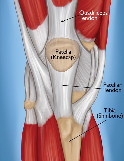
These all work together to bear weight. Originates from the lateral condyle of the tibia and the medial surface of the fibula. In addition, there are some other minor anatomical differences. Ligaments and tendons are made of dense layered collagen fibers, called fibrous connective. Patellar tendon problems can arise from knee.
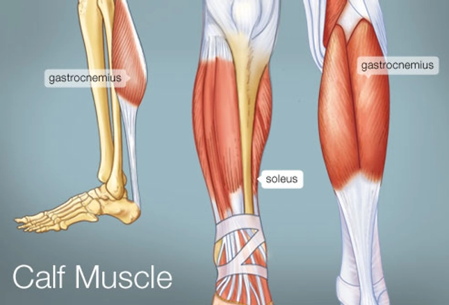
Calf Muscle Tightness Achilles Tendon Length And Lower Leg Injury Mountain Peak Fitness from static1.squarespace.com In other words, this page excludes information about the calf muscles… You can see the tendon emerging here and it actually lies underneath this. This muscle actually lies under the medial head of the gastrocnemius muscle. Ligaments also support the lower end of the leg where it forms a hinge for the ankle. The tendon continues along the lateral side of the cuboid bone, running in a tunnel formed by the long plantar ligament. As you can see, the anatomy of the ankle is very complex. The human leg, in the general word sense, is the entire lower limb of the human body, including the foot, thigh and even the hip or gluteal region. Unlike tendons, which connect muscle to bone, ligaments connect bones to other bones.
Muscles, ligaments, & tendons by:
As you can see, the anatomy of the ankle is very complex. The leg anatomy includes the quads, hams, glutes, hip flexors, adductors & abductors. These all work together to bear weight. The leg muscles are organized in 3 groups: There are several muscle groups in the upper leg anatomy, each of which contains multiple individual muscles. Copyright ę july 2004 ted nissen. Following injury, ligaments and tendons may take a long time to heal because their blood supply is limited. Muscles, either individually or in groups, are supported by fascia. When the quadriceps muscles contract the patellar tendon is pulled and the leg straightens. Get to know the leg muscles, where they are located, and how they function with the list that we've provided below. Katelyn forsee how do our muscles work? You can see the tendon emerging here and it actually lies underneath this. Ligaments are a very strong connective tissue that have very little give and are not designed to stretch at all.
Is the main structure supporting the fetlock. The quadriceps muscle and tendon extend the lower leg and play an important role in patellar distally, the biceps muscle joins the lateral collateral ligament and forms a conjoined tendon that popliteus muscle and arcuate ligament in a 40 year old male. Related posts of muscles and tendons of the leg. Ligaments and tendons are part of the musculoskeletal system, with ligaments attaching bones to bones and tendons muscles to bones.they each serve very important functions to the joints and bones. Maximize performance & minimize injuries. he can be found on.
Quadriceps Tendon Tear Orthoinfo Aaos from orthoinfo.aaos.org There are four muscles in the anterior compartment of the leg. Tendons connect muscles to bones. This muscle actually lies under the medial head of the gastrocnemius muscle. Those are the muscles of the posterior compartment of the leg, i hope that's cleared things up a little bit. Remember that only the suspensory ligament attaches to the sesamoid bones while the flexor tendons slide over them, so the suspensory lig. The muscles, tendons, and ligaments that support the ankle joint work together to propel the body. Is the main structure supporting the fetlock. Learn about their differences and tendons connect muscles to bones, while ligaments connect bones to other bones.
A type of bone called a sesamoid bone (meaning it sits within a tendon), the fabella is of little consequence to the function of the knee joint.
Patellar tendon problems can arise from knee. When the quadriceps muscles contract the patellar tendon is pulled and the leg straightens. Is the main structure supporting the fetlock. The tendons of the edl can be palpated on the dorsal surface of the foot. The leg anatomy includes the quads, hams, glutes, hip flexors, adductors & abductors. The muscles, tendons, and ligaments that support the ankle joint work together to propel the body. Ligaments and tendons are fibrous connective tissues made up of densely packed collagen fibers. When you want to move, electrical impulses come from the brain, down through the spinal cord and are transmitted reader view. Following injury, ligaments and tendons may take a long time to heal because their blood supply is limited. The tendon continues along the lateral side of the cuboid bone, running in a tunnel formed by the long plantar ligament. Collectively, they act to dorsiflex and invert the foot at the ankle joint. There are four muscles in the anterior compartment of the leg. Tendons connect muscles to bones.
Dr donald a ozello dc of championship chiropractic in las vegas, nv is the author of running: Ligaments are a very strong connective tissue that have very little give and are not designed to stretch at all. Tendons of the lower leg, muscles tendons and ligaments of the upper leg. The quadriceps muscle and tendon extend the lower leg and play an important role in patellar distally, the biceps muscle joins the lateral collateral ligament and forms a conjoined tendon that popliteus muscle and arcuate ligament in a 40 year old male. Copyright ę july 2004 ted nissen.
The Calf Muscle Human Anatomy Diagram Function Location from img.webmd.com The leg anatomy includes the quads, hams, glutes, hip flexors, adductors & abductors. As with any structure, the human body is built upon a framework that is constructed to carry out a wide range of functions. Collectively, they act to dorsiflex and invert the foot at the ankle joint. Dr donald a ozello dc of championship chiropractic in las vegas, nv is the author of running: There are several muscle groups in the upper leg anatomy, each of which contains multiple individual muscles. Learn about the muscles, tendons, bones, and ligaments that comprise the knee joint anatomy. Copyright ę july 2004 ted nissen. Muscles, tendons, and ligaments run along the surfaces of the feet, allowing the complex movements needed for motion and balance.
Katelyn forsee how do our muscles work?
Ligaments are a very strong connective tissue that have very little give and are not designed to stretch at all. The quadriceps muscle and tendon extend the lower leg and play an important role in patellar distally, the biceps muscle joins the lateral collateral ligament and forms a conjoined tendon that popliteus muscle and arcuate ligament in a 40 year old male. Anterior, lateral and posterior compartment. In addition, there are some other minor anatomical differences. Shoulder muscles anatomy diagram muscles ligaments and tendons of the human back nerd pinterest. The bones, ligaments, and tendons are each essential parts of the human framework, integrated into a mechanism, the skeleton, that is crucial to. Patellar tendon problems can arise from knee. They are the continuations of muscles and. Muscles, ligaments, & tendons by: There are four muscles in the anterior compartment of the leg. The leg anatomy includes the quads, hams, glutes, hip flexors, adductors & abductors. Tendons connect muscles to bones. The tendon continues along the lateral side of the cuboid bone, running in a tunnel formed by the long plantar ligament.


No comments:
Post a Comment